WE NEED
MORE TEETH
The Tyrannosaurus rex casts at the Sedgwick Museum
By the Sedgwick Museum of Earth Sciences


An exciting new collection of dinosaur casts was donated to the Sedgwick Museum in 2017. Among them, those of a prehistoric icon: Tyrannosaurus rex.
Eye surgeon Dr. Andrew Hempel bequeathed his
large collection of dinosaur skull and bone replicas to the museum in the summer of 2017.

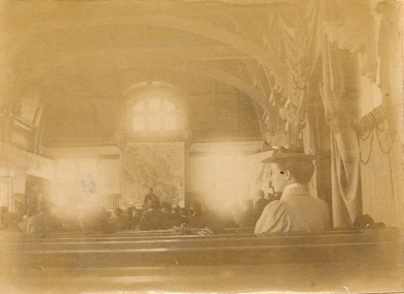
An image from the original display in the museum
An image from the original display in the museum
It focuses on the skull of a Tyrannosaurus rex nicknamed ‘Stan’. This online exhibition also explains why and how replicas or casts are made of fossils. There is also a look behind-the-scenes into the curation of collections. This is a key part of our work at the museum.
Tyrannosaurus rex factfile
Name: Tyrannosaurus rex (Tyrant lizard king).
Age: Late Cretaceous (Around 66 Million Years ago) .
Location: North America.
Rock: Hell Creek and Lance Formations.
Length: Between 12–13.3 metres.
Height: Up to 3.7 meters at the hip.
Diet: Other dinosaurs, including Triceratops and Edmontosaurus.
Habitat: Subtropical floodplains and coastal plains at the edge of a large shallow sea.
First discovered: 1902 by American palaeontologist Barnum ‘Mr Bones’ Brown.
Named: 1905 by American palaeontologist Henry Fairfield Osborn.
Number of teeth: Around 50.


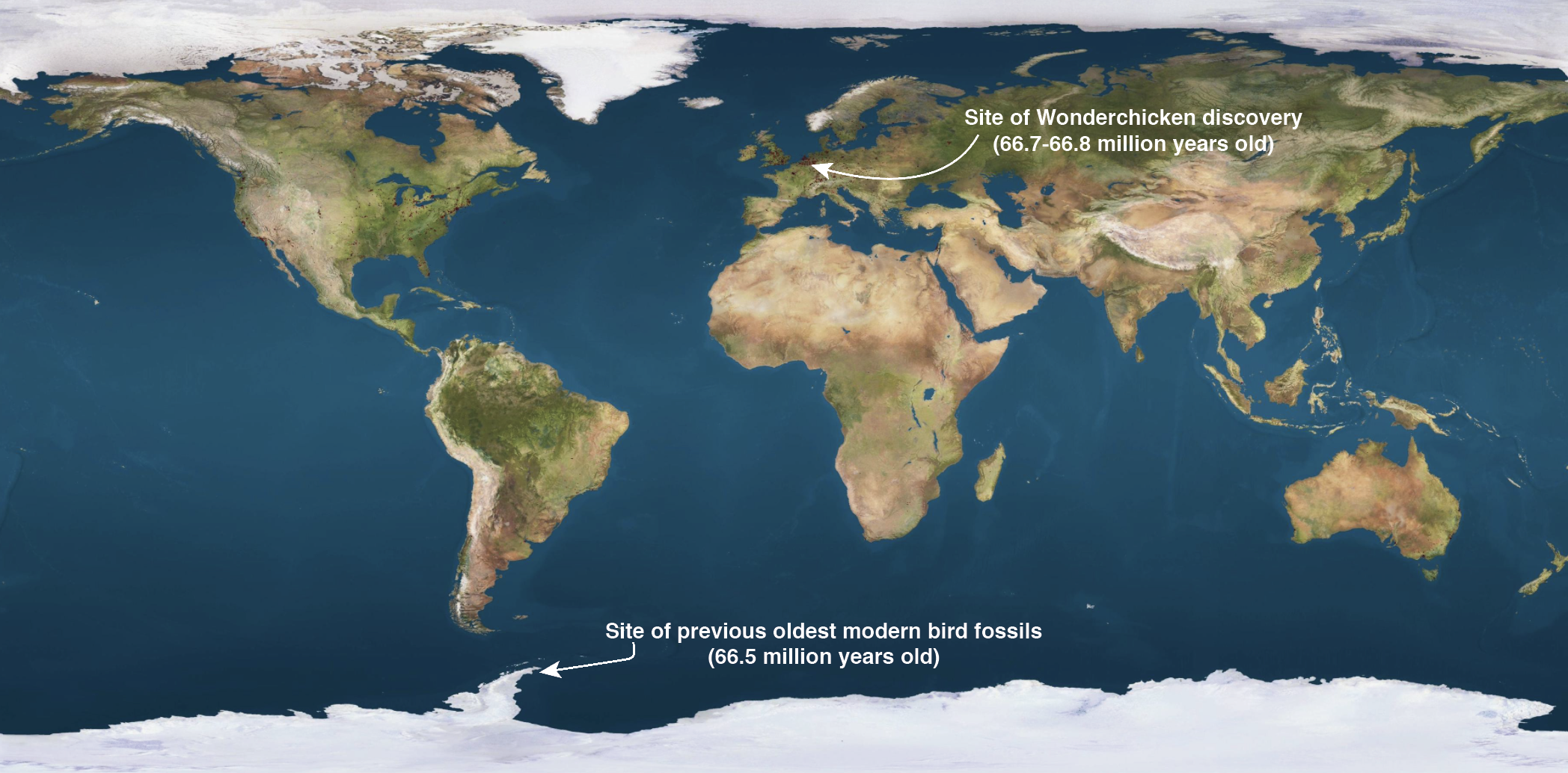
Where did it live?
Tyrannosaurus rex remains have only been found in Western North America. Most come from the Badlands of South Dakota
and Montana, USA, but have been found as far south as Texas.
T. rex remains have also been unearthed in Alberta and
Saskatchewan, Canada.

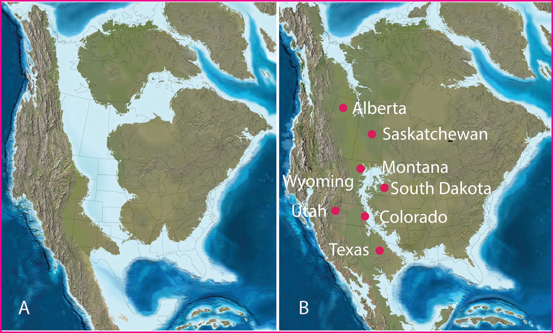
Paleogeographic maps of North America during the late Creataceous.
(A) late Campanian (c. 75 Million Years Ago) (B) late Maastrichtian (c. 66 Million Years Ago), when T. rex lived.
Paleogeographic maps of North America during the late Creataceous.
(A) late Campanian (c. 75 Million Years Ago) (B) late Maastrichtian (c. 66 Million Years Ago), when T. rex lived.
Tyrannosaurus rex’s ancestors lived in Asia. They migrated across a land-bridge between Asia and western North America during the
Cretaceous Period. A large sea split North America in two for much of the Cretaceous. This may be why no T. rex remains have been found in eastern North America.
Changing ideas

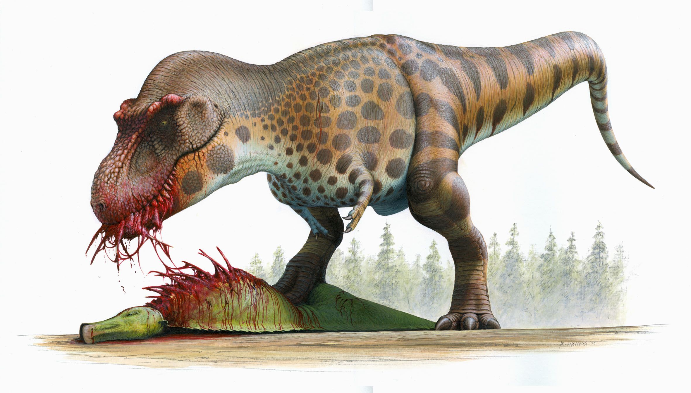
Credit: Robert Nicholls 2009
Credit: Robert Nicholls 2009
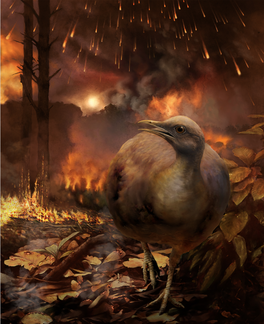
Palaeoartist Bob Nicholls painted this image for the museum in 2009. It was accurate at the time. New theories and discoveries in the 10 years since it was painted have changed our understanding of how T. rex looked. This has meant that for several years it was an out-dated reconstruction, but we know now that it is still quite accurate. Museums work hard to keep up with new research, but old ideas and reconstructions still often remain on display.

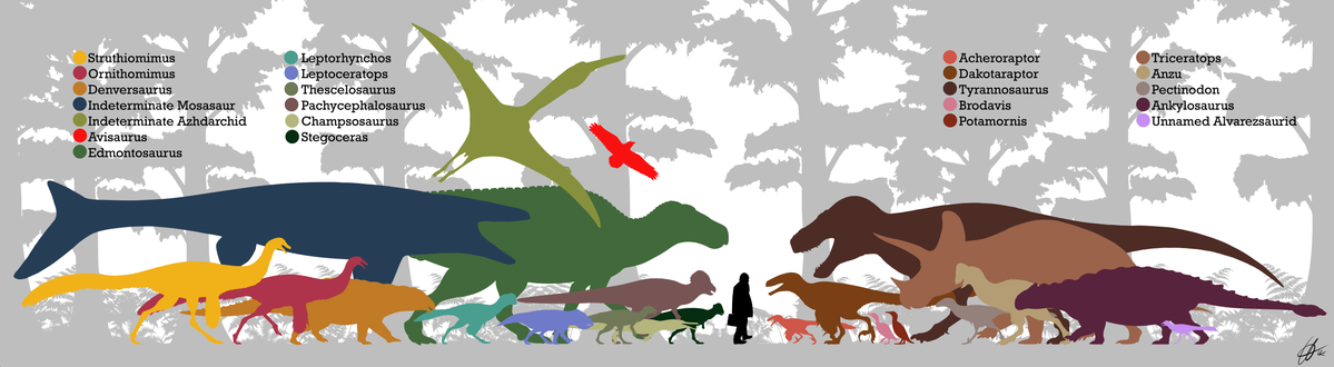
Dinosaurs, pterosaurs and some marine reptiles discovered in the Hell Creek Formation. Image by PaleoNeolitic, CC BY-SA 4.0, via Wikimedia Commons
Dinosaurs, pterosaurs and some marine reptiles discovered in the Hell Creek Formation. Image by PaleoNeolitic, CC BY-SA 4.0, via Wikimedia Commons
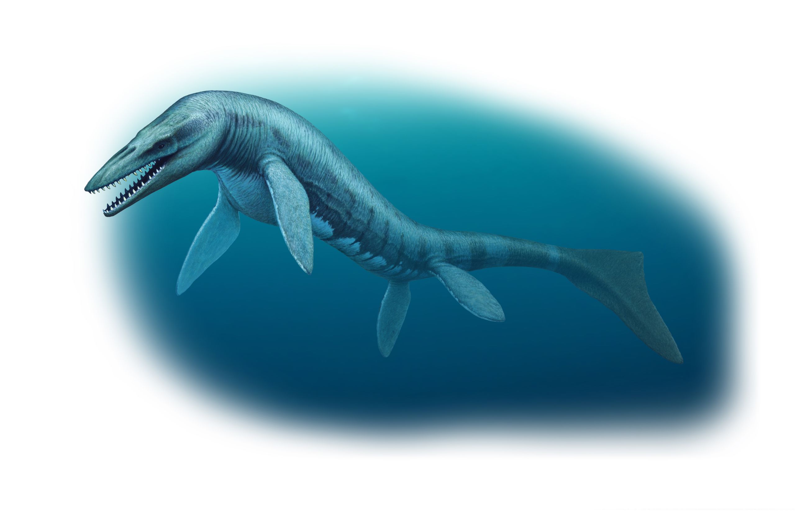
“KING OF THE TRYANTS”
Tyrannosaurus rex was one of the largest theropod dinosaurs, but its ancestors were much smaller and were feathered. Its closest
living relatives are also much smaller and are feathered - they are birds.
The word theropod means “beast foot” in Greek.
Theropod dinosaurs all had hollow bones and three-toed feet. Tyrannosaurus rex is from a diverse group of theropod dinosaurs called the coelurosaurs. In this group are the largest and smallest theropods, as well as plant-eating ones. Using fossil evidence, many palaeontologists think that most coelurosaurs had coats of primitive feathers. Birds evolved from small, feathered coelurosaurs, making them living dinosaurs.
Dinosaur family trees
Theropod and sauropod dinosaurs are traditionally grouped together as saurischian dinosaurs.

In the late 1880s, palaeontologist Harry Seeley divided dinosaurs into the two orders, Saurischia and Ornithischia, based on their hip structures.
In the late 1880s, palaeontologist Harry Seeley divided dinosaurs into the two orders, Saurischia and Ornithischia, based on their hip structures.
A 2017 study by Cambridge palaeontologists proposed a new dinosaur tree. Their study found theropods were more closely related to ornithischian dinosaurs. They gave this new group the name Ornithoscelida.

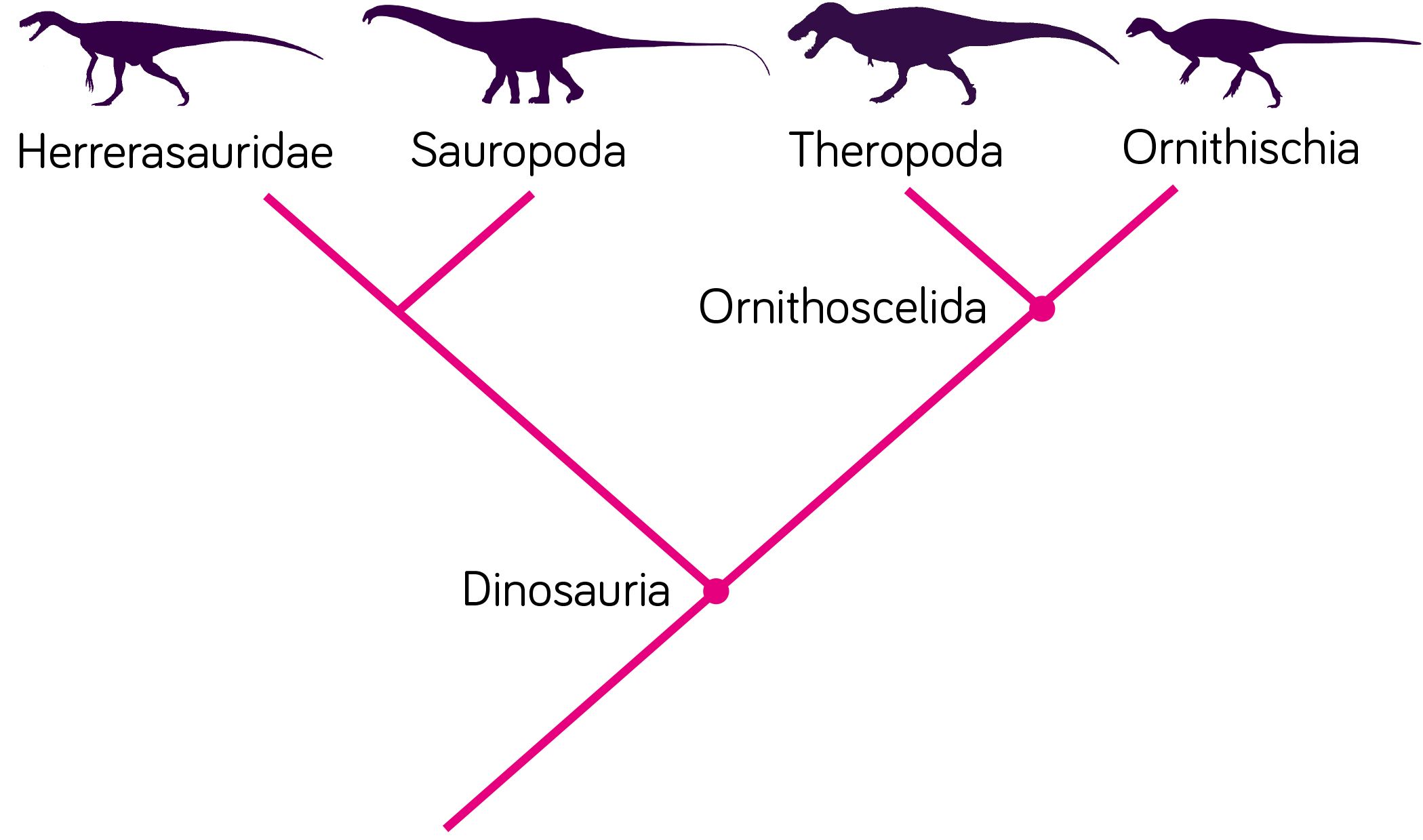
The new dinosaur tree, proposed by Palaeontologists Matthew Baron, David Norman and Paul Barrett
The new dinosaur tree, proposed by Palaeontologists Matthew Baron, David Norman and Paul Barrett
“Big things have small beginnings”
Tyrannosaurus rex evolved from much smaller ancestors.
Many of these had feather-like coverings on their bodies. Some also had feathers. Because of this, some palaeontologists think T. rex also had bristly feather-like covering when it was young, although no direct evidence has been found.

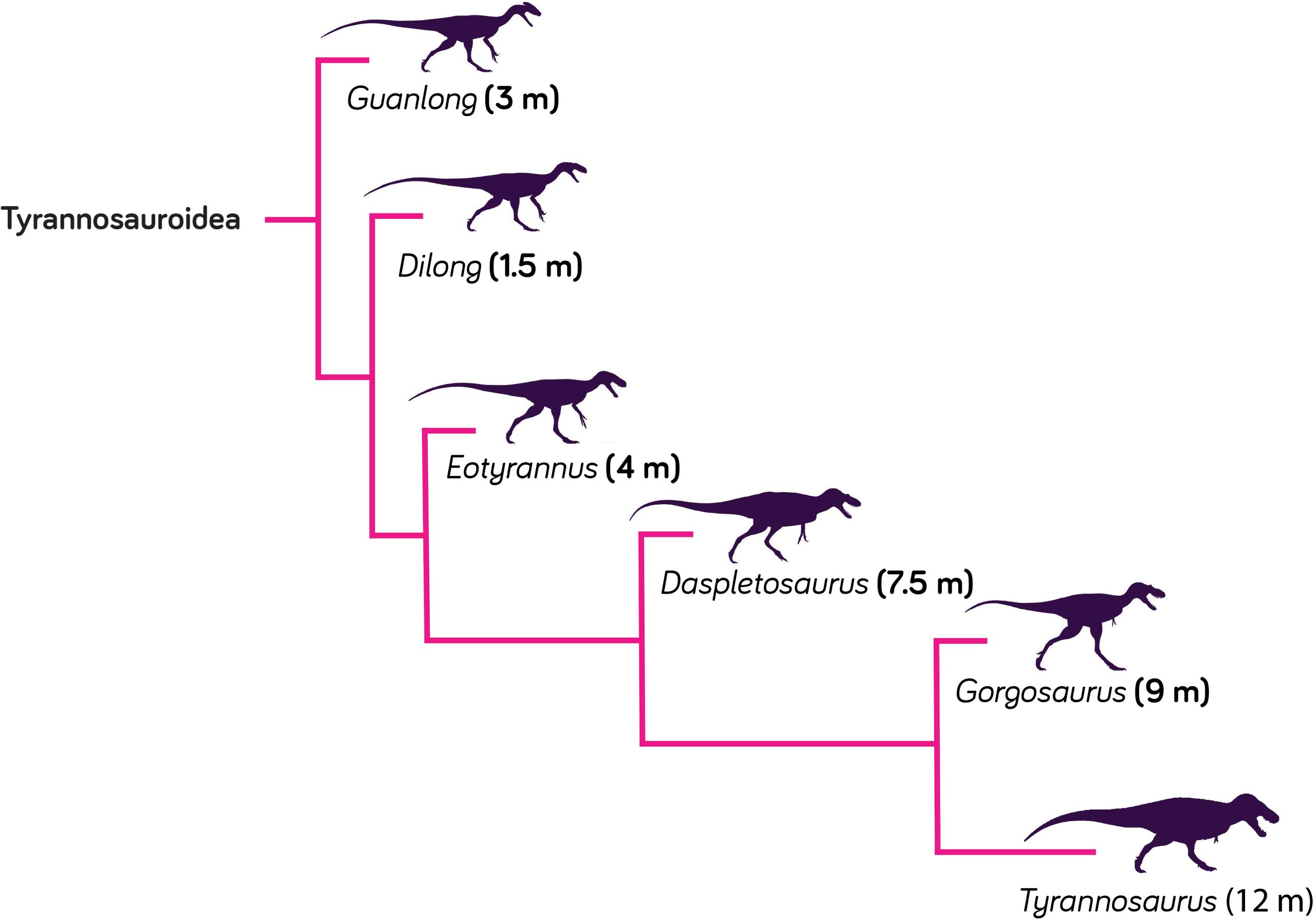
Tyrannosaurus rex's early ancestors, like Guanlong and Dilong, were discovered in China. They were much smaller than T. rex and had feather-like coverings.
Tyrannosaurus rex's early ancestors, like Guanlong and Dilong, were discovered in China. They were much smaller than T. rex and had feather-like coverings.
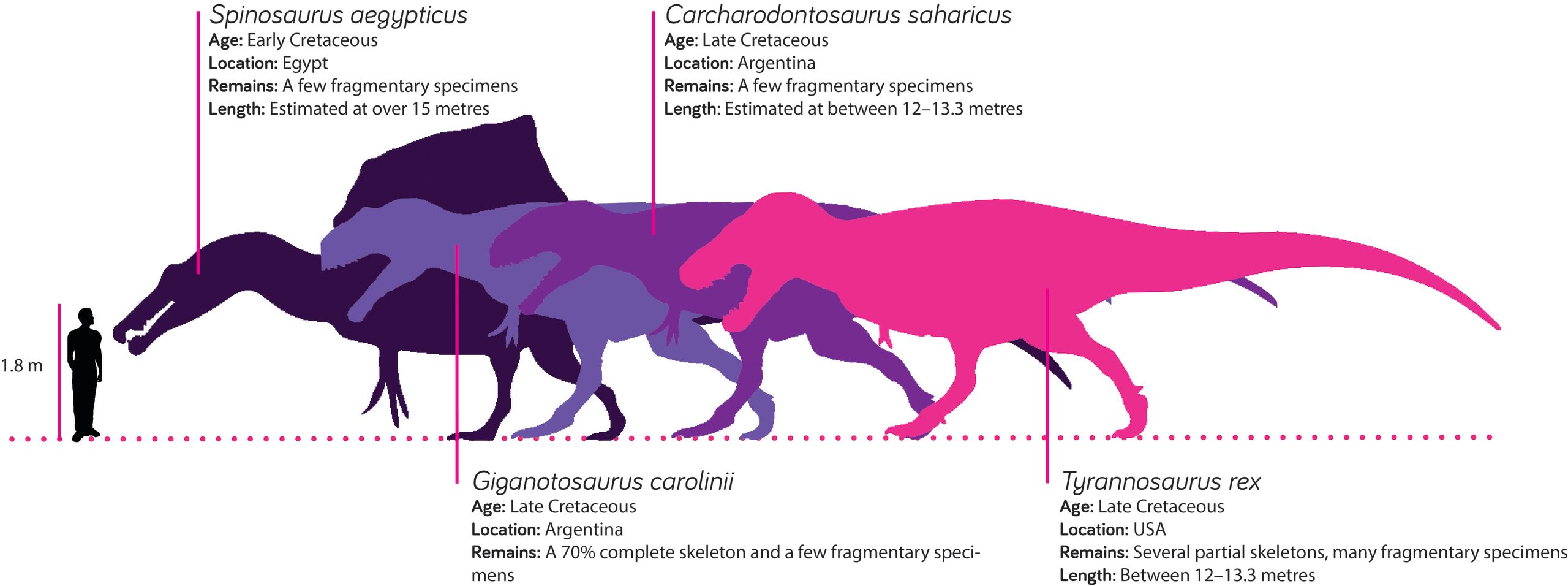
Still the king?
For many years Tyrannosaurus rex was the biggest theropod dinosaur known. But other giants have been discovered in
Africa and South America that are close to, or even larger than, T. rex. Fossils of these huge predators are rare and incomplete, so their sizes are still debated by palaeontologists.

BHI 3033 - ‘STAN’
Nicknamed to honour amateur paleontologist Stan Sacrison, ‘Stan’ is one of the most complete and well-known Tyrannosaurus rex specimens ever discovered.
Stan the T. rex was found in the Hell Creek Formation near Buffalo, South Dakota, in the spring of 1987.
Collected in 1992, it was first thought to be a Triceratops skeleton. Over 50 Tyrannosaurus rex specimens have been found since it was first named in 1905. More complete skeletons, like Stan’s, are rare. This makes them valuable to palaeontologists, allowing them to learn a huge amount about T. rex. Several casts of Stan’s skull are in Dr Hempel’s collection.
How big was ‘Stan’?
At a little over 11 metres long, Stan was not fully-grown when it died. Palaeontologists estimate that it was around 18 years old. The largest, oldest Tyrannosaurus rex discovered is nicknamed ‘Sue’. At 12.3 metres long, it is estimated to be around 30 years old when it died.

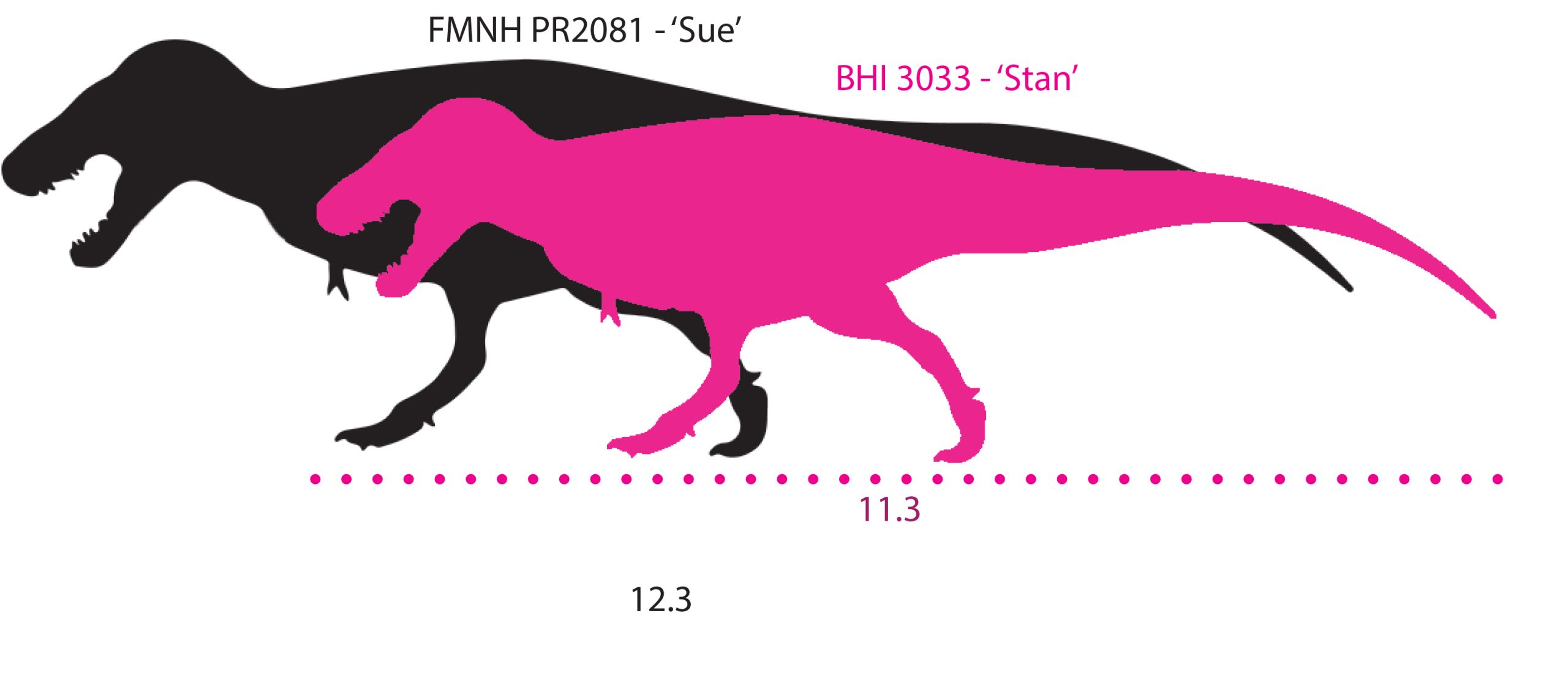
Size comparison of Stan and Sue. Stan is one meter shorter than Sue, and has a more lightly built skeleton
Size comparison of Stan and Sue. Stan is one meter shorter than Sue, and has a more lightly built skeleton
Puzzle pieces
Stan is one of the most complete Tyrannosaurus rex
skeletons discovered. 199 of its bones have been found, which is around 63% of the total skeleton. Stan is missing both arms, several ribs and some tail and foot bones. Part of Stan’s pelvis and its gastralia, or belly ribs, are also missing.

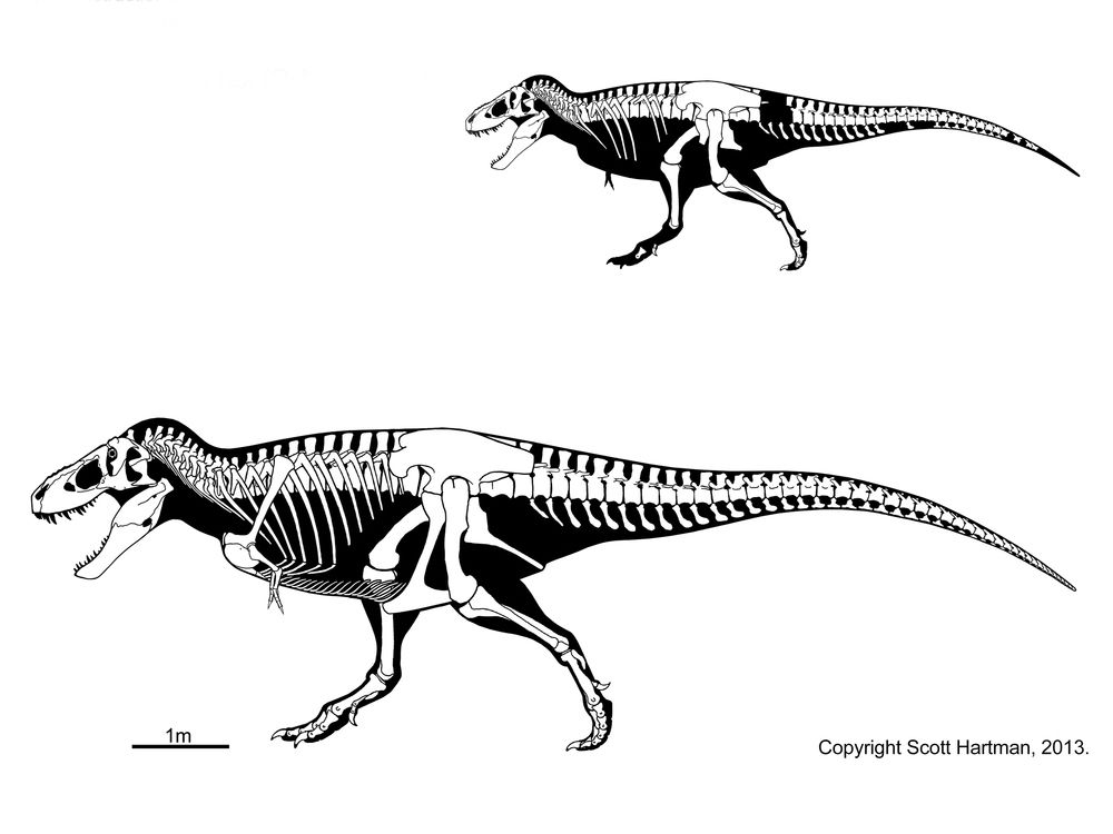
Diagram of the known remains and complete skeleton of 'Stan' Copyright Scott Hartman 2013 https://www.skeletaldrawing.com/theropods/tyrannosaurus-stan
Diagram of the known remains and complete skeleton of 'Stan' Copyright Scott Hartman 2013 https://www.skeletaldrawing.com/theropods/tyrannosaurus-stan
Boy or girl?
Female and male animals can often be told apart by their different sizes, shapes, colours and markings. This is called sexual dimorphism. It can difficult to see in fossils. Stan’s lightly-built skeleton was once thought to mean it was a male T. rex, but now many palaeontologists are not so sure.
STAN’S
SKULL
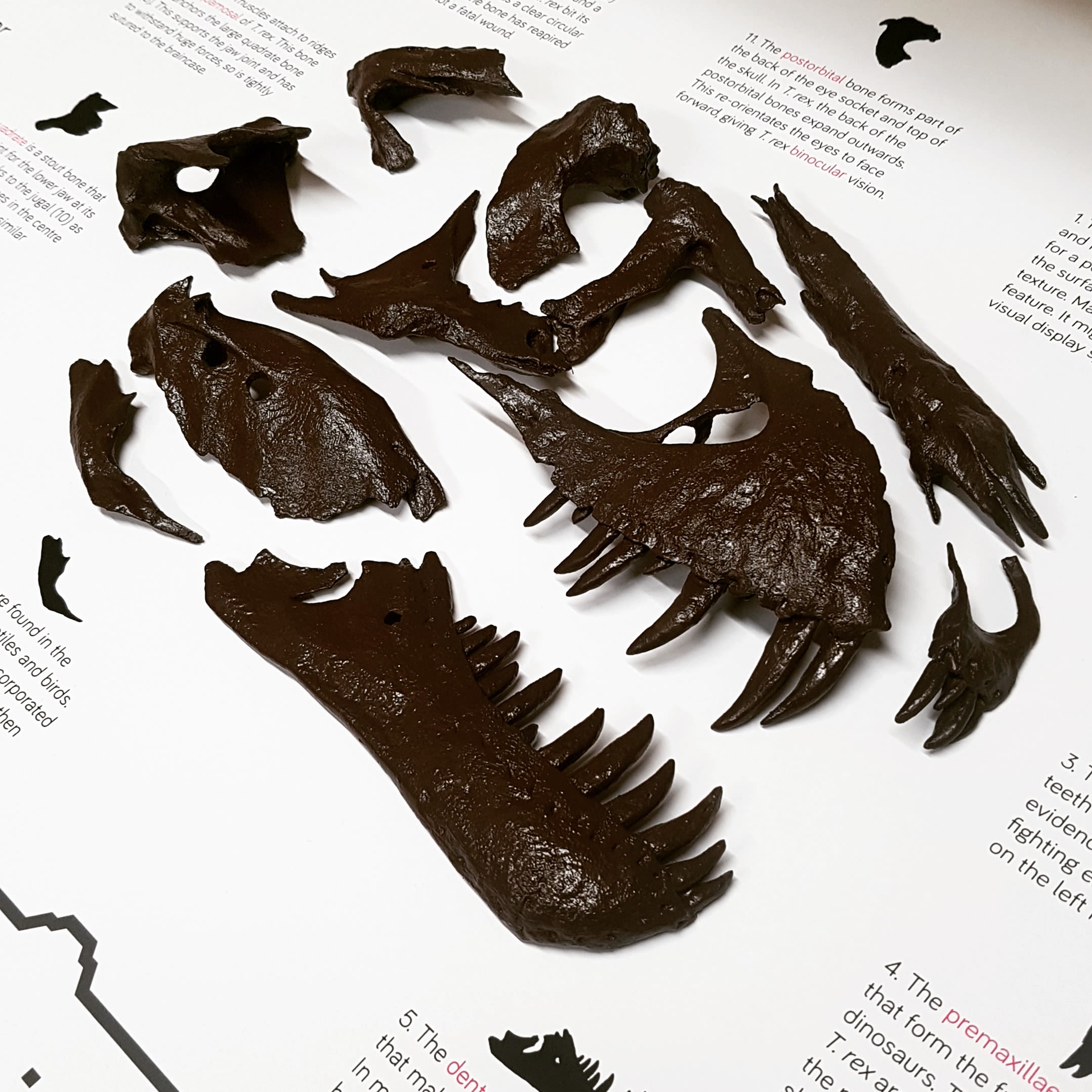
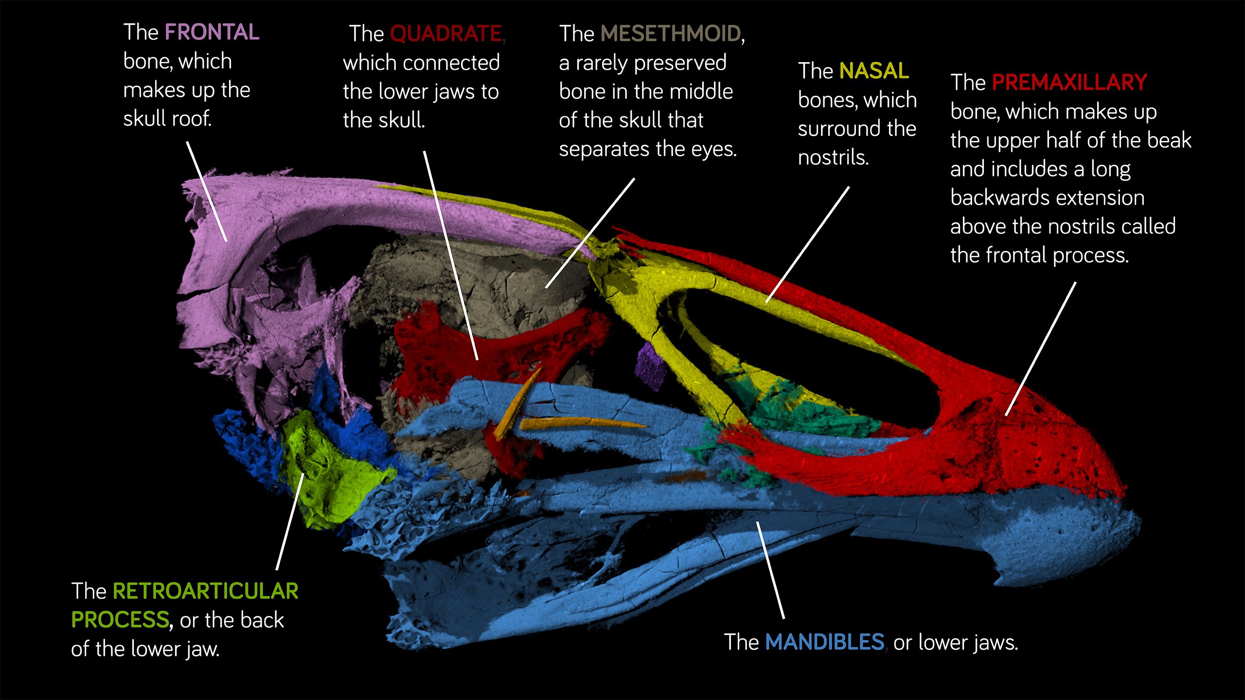
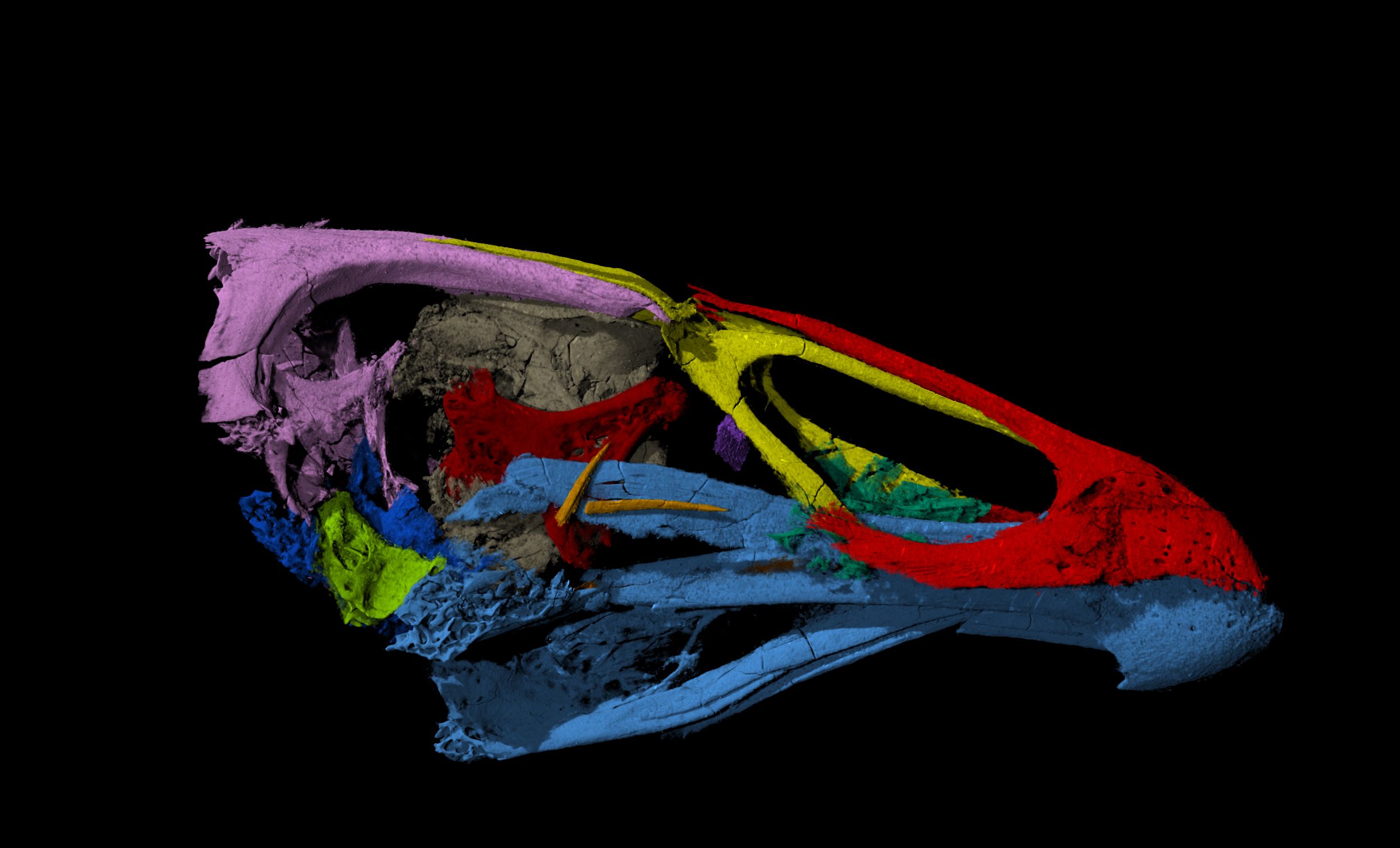
At 1.4 m long, Stan’s huge skull is over 10% of its total body length. It is one of the most complete Tyrannosaurus rex skulls ever found.
47 of Stan’s skull bones were collected. The skull bones are preserved separately, meaning palaeontologists can study each bone in careful detail. A 3-D scan of Stan’s skull has been used to measure its bite force. It has an estimated bite force of 6.8 tonnes, which is the largest of any animal, living or extinct.

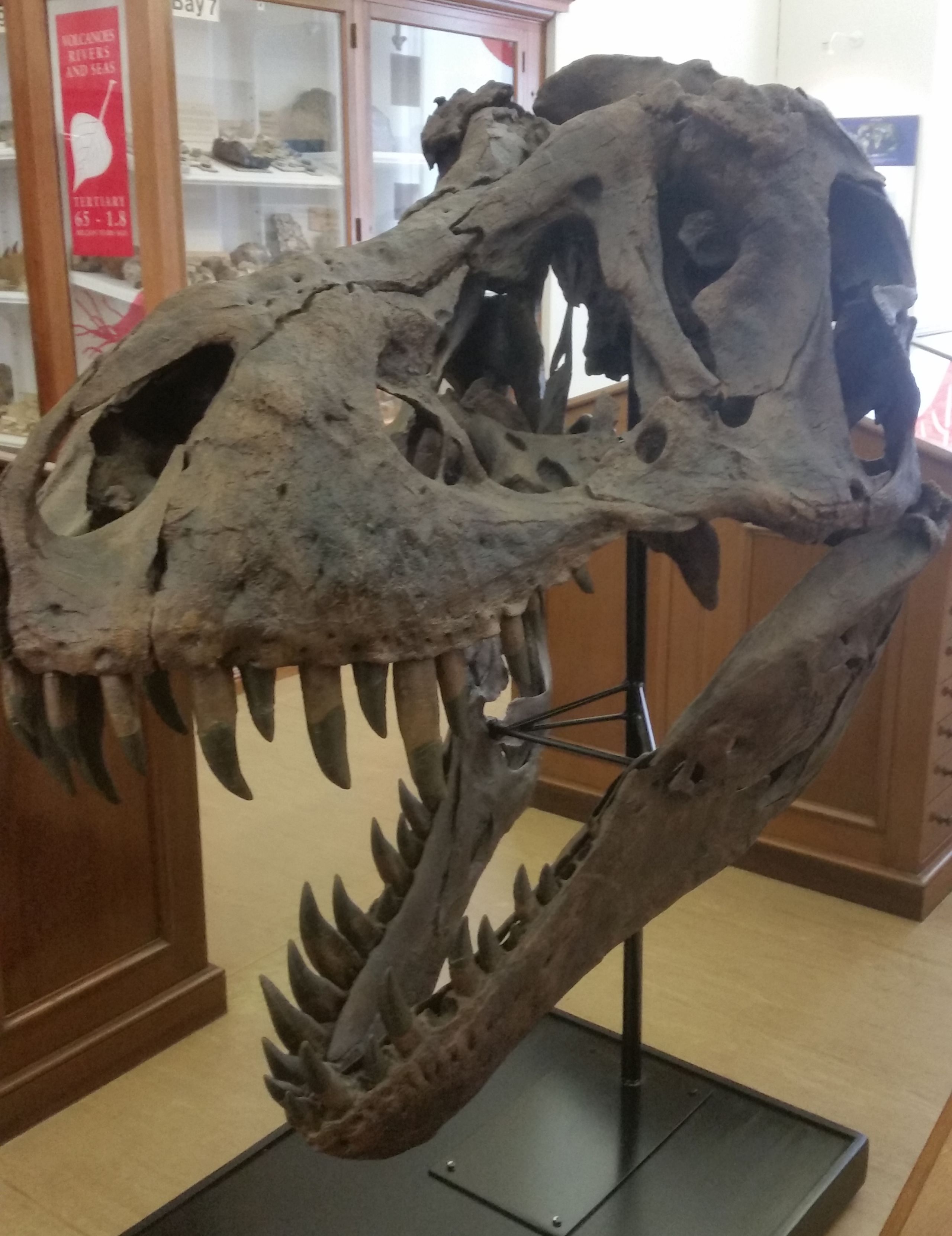
The cast of 'Stan''s skull on display in the museum
The cast of 'Stan''s skull on display in the museum
Many of the bones of Stan’s skull have holes in them. The name for these holes is fenestra, which means ‘window’ in
Latin. The fenestrae in the skulls of theropods, such as T. rex, helped reduce their weight and provided
attachments for muscles. Stan’s skull also has several holes which are not fenestrae. These are healed injuries, which are called pathologies. We know they are injuries as the holes do not match on either side of the skull.
Dino damage
The irregular holes on Stan’s cheek and lower jaw bones are healed injuries. They were inflicted on Stan whilst he was still alive. Many Tyrannosaurus rex specimens have pathologies
on their skulls and skeleton. These tell us that violence was a routine part of T. rex’s life.

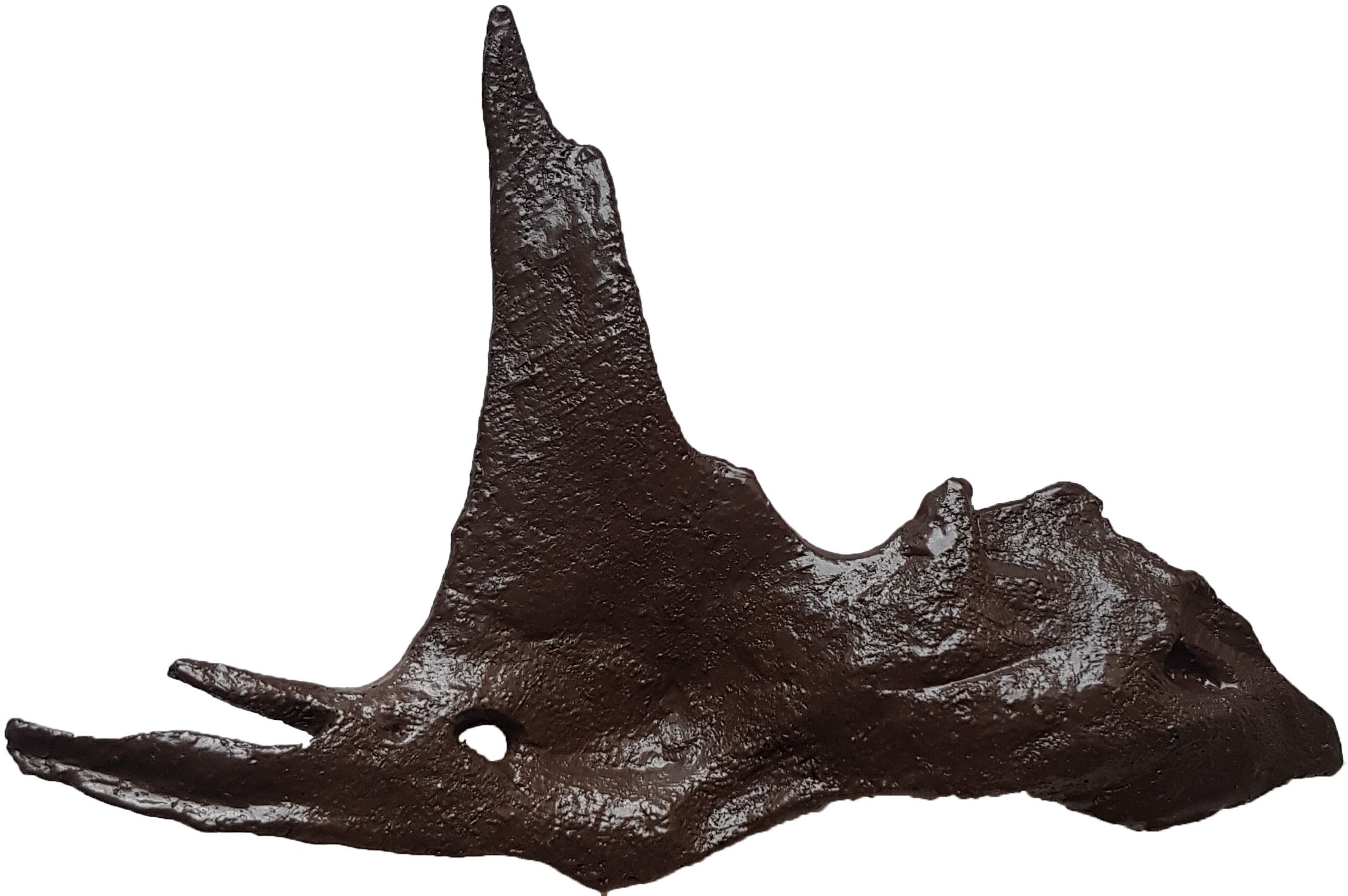
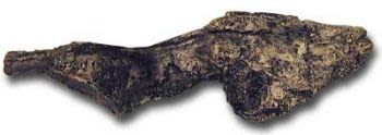
A cast of 'Stan's' jugal, or cheekbone. The hole in this bone was made by another Tyrannosaurus rex whilst Stan was alive.
A cast of 'Stan's' jugal, or cheekbone. The hole in this bone was made by another Tyrannosaurus rex whilst Stan was alive.
The bones at the back of Stan’s skull, which house the brain, show an unusual pathology. Part of the crest on the left-hand side is missing, and there is a round hole below this. This hole matches the shape of a tooth from the lower jaw of another Tyrannosaurus rex.

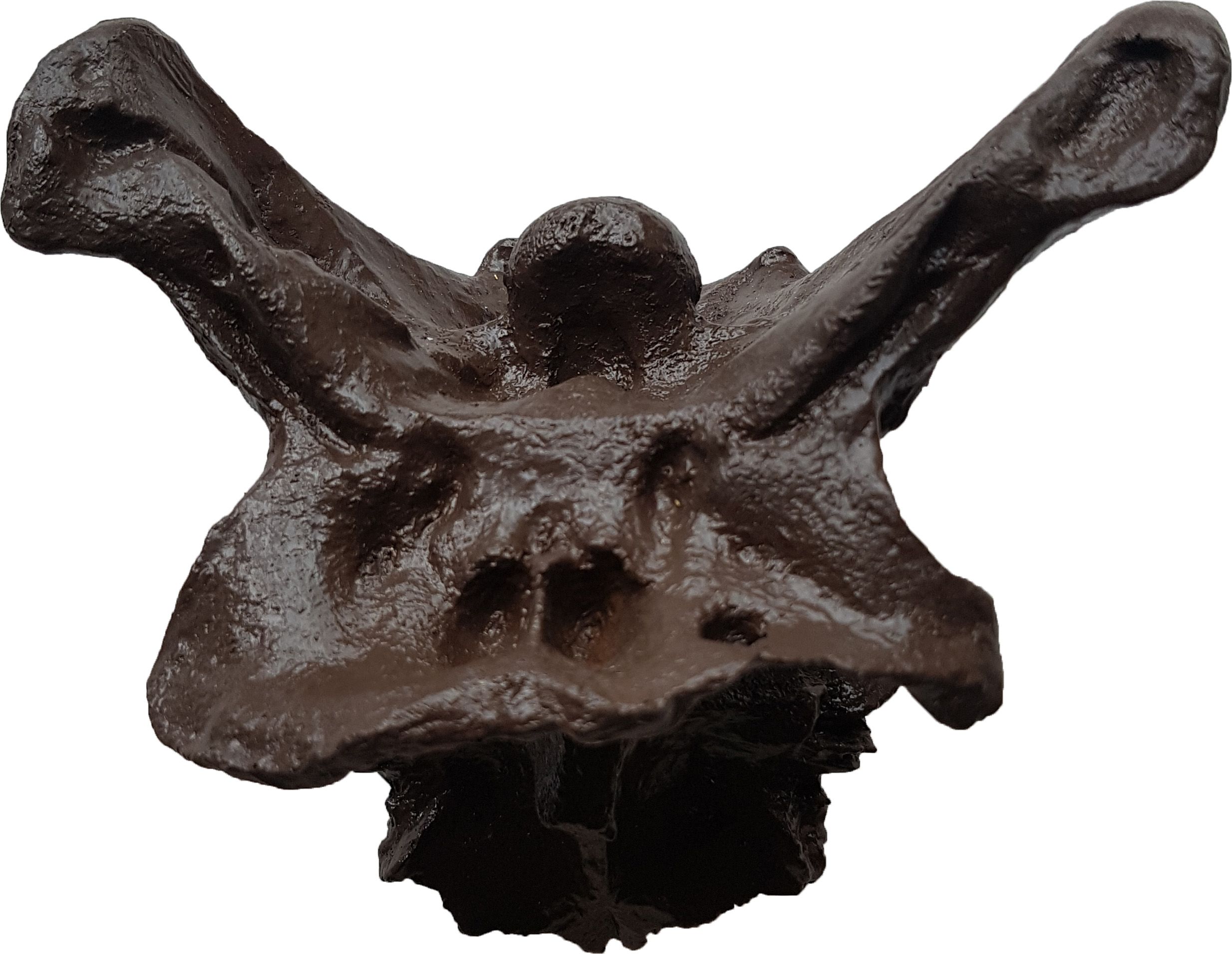
A cast of bones at the back of 'Stan's' skull. The hole in this bone was made by another Tyrannosaurus rex whilst Stan was alive.
A cast of bones at the back of 'Stan's' skull. The hole in this bone was made by another Tyrannosaurus rex whilst Stan was alive.
Stan’s pathologies are not found only on his skull. Its skeleton also has several broken and healed ribs, and broken neck
vertebrae. Two neck vertebrae have fused together as they healed. Extra bone has grown on a third neck vertebrae,
obstructing its movement. Its hips and tail also show damage.
‘ALL TEETH, NO BRAINS?’
Tyrannosaurus rex is often portrayed as a mindless killer. It had a huge skull and teeth, but a brain tiny in comparison to its body size. So, was T. rex all teeth and no brains?
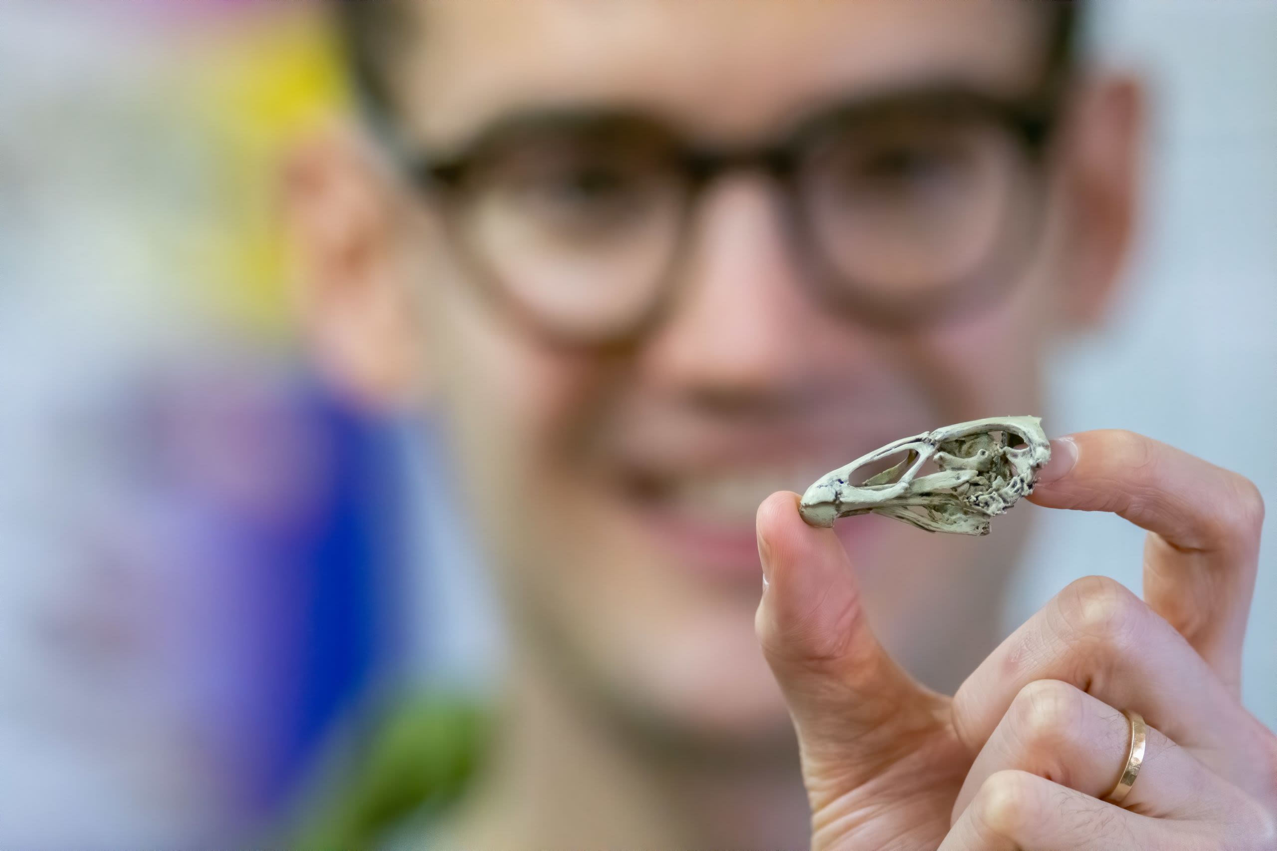
Well preserved T. rex skulls, like Stan’s, allow palaeontologists to study an important part of its body: its brain. They can make casts of T. rex braincases, called
endocasts, by scanning the bones. Palaeontologists can compare the structure of a T. rex brain with its
living relatives, crocodiles and birds. This helps them to learn about its senses. Theories about T. rex behaviour are made using a combination of research on the skeleton and casts of its brain.
Brain cast

This is a replica of the endocranial cavity, or braincase, of
T. rex. Despite its small size, it is quite large and advanced for a dinosaur brain. Birds and reptiles are the closest living animals that can be used to compare brain size. T. rex has a ‘brain-to-body’ size ratio that is more similar to an alligator than a bird.
Super senses


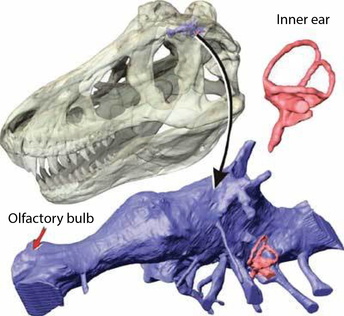
CT scans of the endocranial cavity of T. rex, in blue, reveal a lot about its senses. The large olfactory bulbs show that it had a strong sense of smell. Long looped tubes in its inner ear, in pink, gave it
excellent balance. The tubes projecting from the endocast are complex nerves to co-ordinate the activities of this animal.
Infrasonic hearing
The shape of Tyrannosaurus rex’s inner ear suggests it could hear infrasonic, or low-frequency, sounds. Infrasonic sounds can travel long distances without dissipating. T. rex is more likely to have made low rumbling calls like a crocodile or elephant than the high-pitched sounds birds use to communicate.
Binocular vision
Tyrannosaurus rex had eyes that face forwards, like humans. This is called binocular vision. Binocular vision meant T. rex could judge the distances. Like modern hunters with this ability, T. rex would be able to work out how far away its prey was when it was hunting.
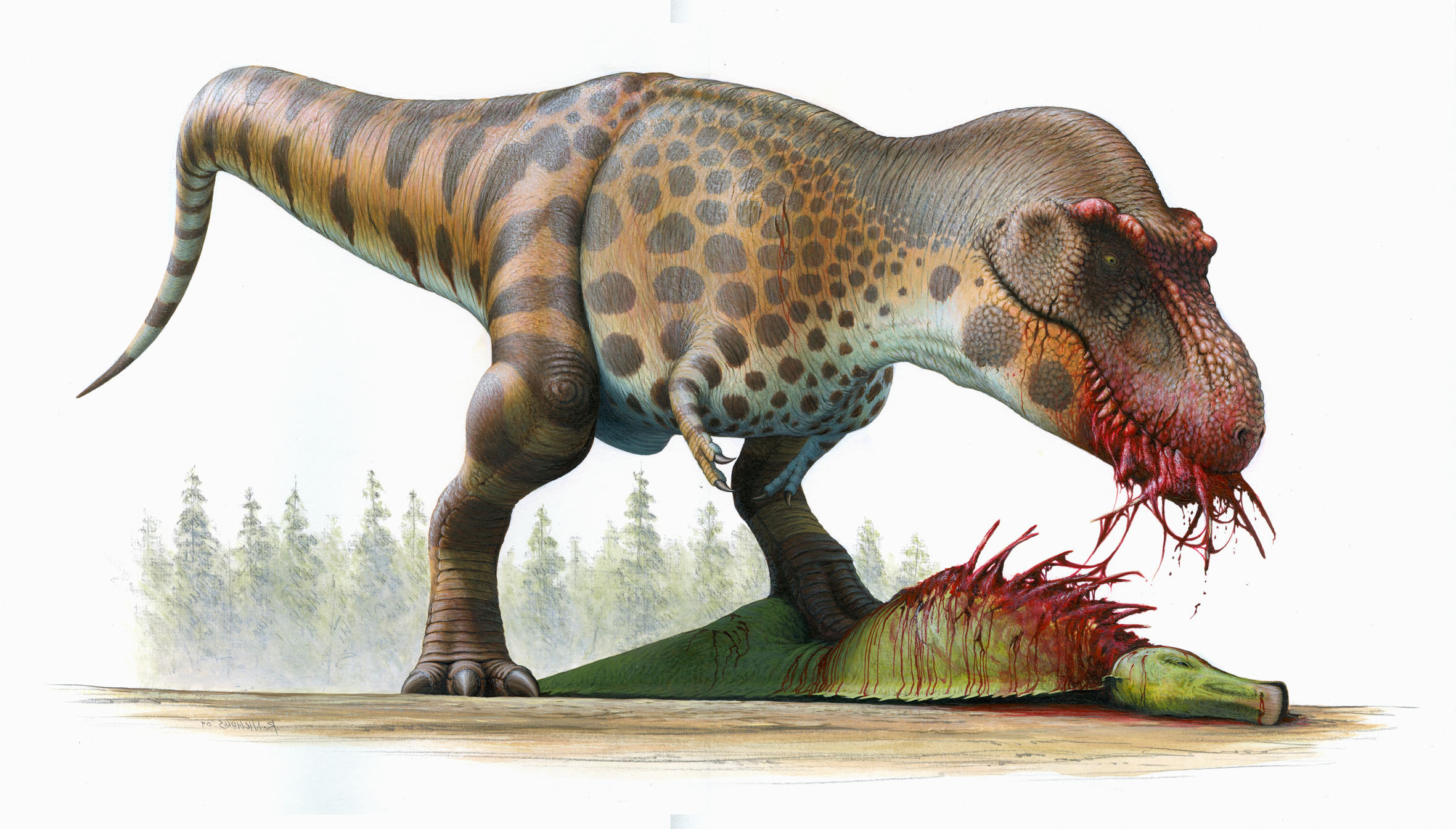
‘My what big teeth you have’

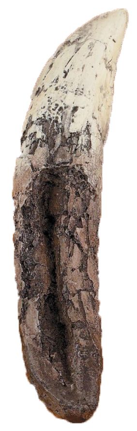
T. rex was a carnivore. Its teeth are the shape of a meat-eater’s and they are the largest of any dinosaur. Each is curved and pointed, with a pair of edges called carinae. Serrations on the carinae, called denticles, helped the teeth to slice flesh. The teeth are very thick, making them strong enough to crack solid bone.
What did it eat?
Sharing the environment with Tyrannosaurus rex were
several other types of dinosaur. The two largest were the
ceratopsian Triceratops, and hadrosaur Edmontosaurus. Healed bite marks on fossils of both dinosaurs are likely to have been made by failed T. rex attacks.
“IS IT
REAL?”
Complete Tyrannosaurus rex skeletons
are rare. Replicas of the best specimens are made so they can be studied and displayed around the world.
Replica fossils allow museums to display specimens they may not otherwise be able to show. They are
often rare or very large, like T. rex skeletons. These
replicas also have scientific value of their own. Stan’s actual skeleton is on display in the Black Hills Institute of Geological Research in Hill City, South Dakota.
The Black Hills Institute produces high quality replicas of many fossils, including Stan. Most of Dr Hempel’s collection of casts are produced by the Institute.
Faithful reproductions

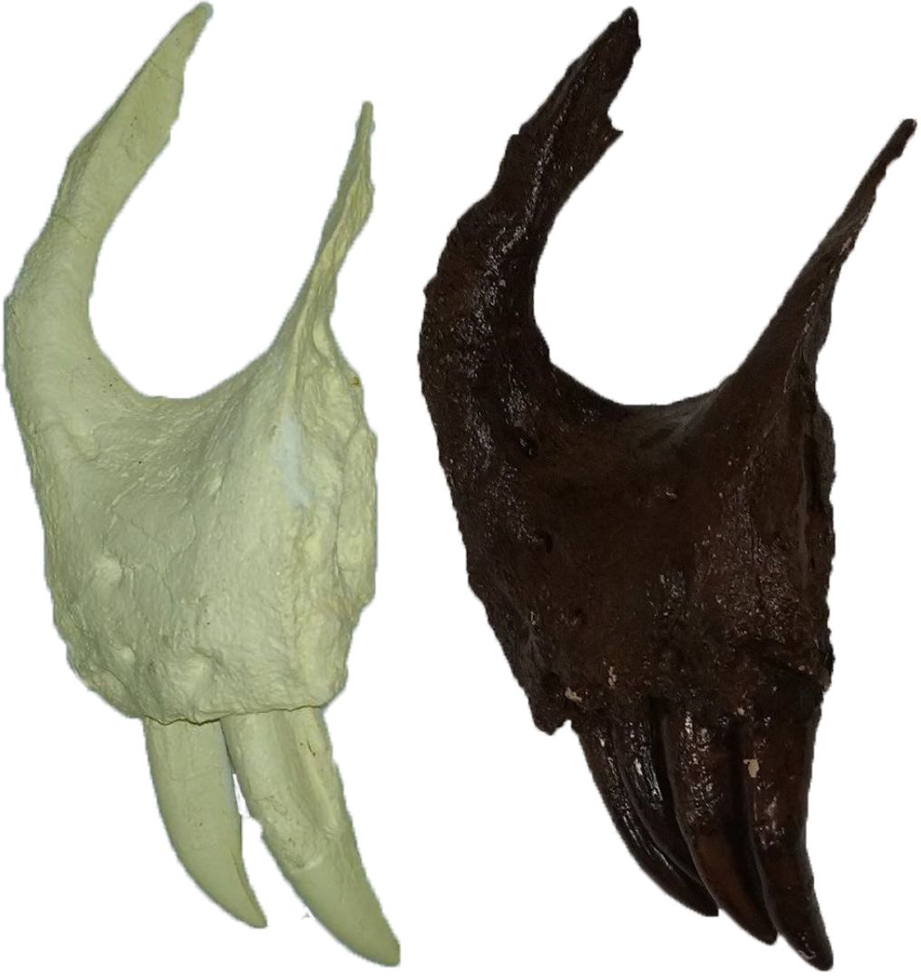
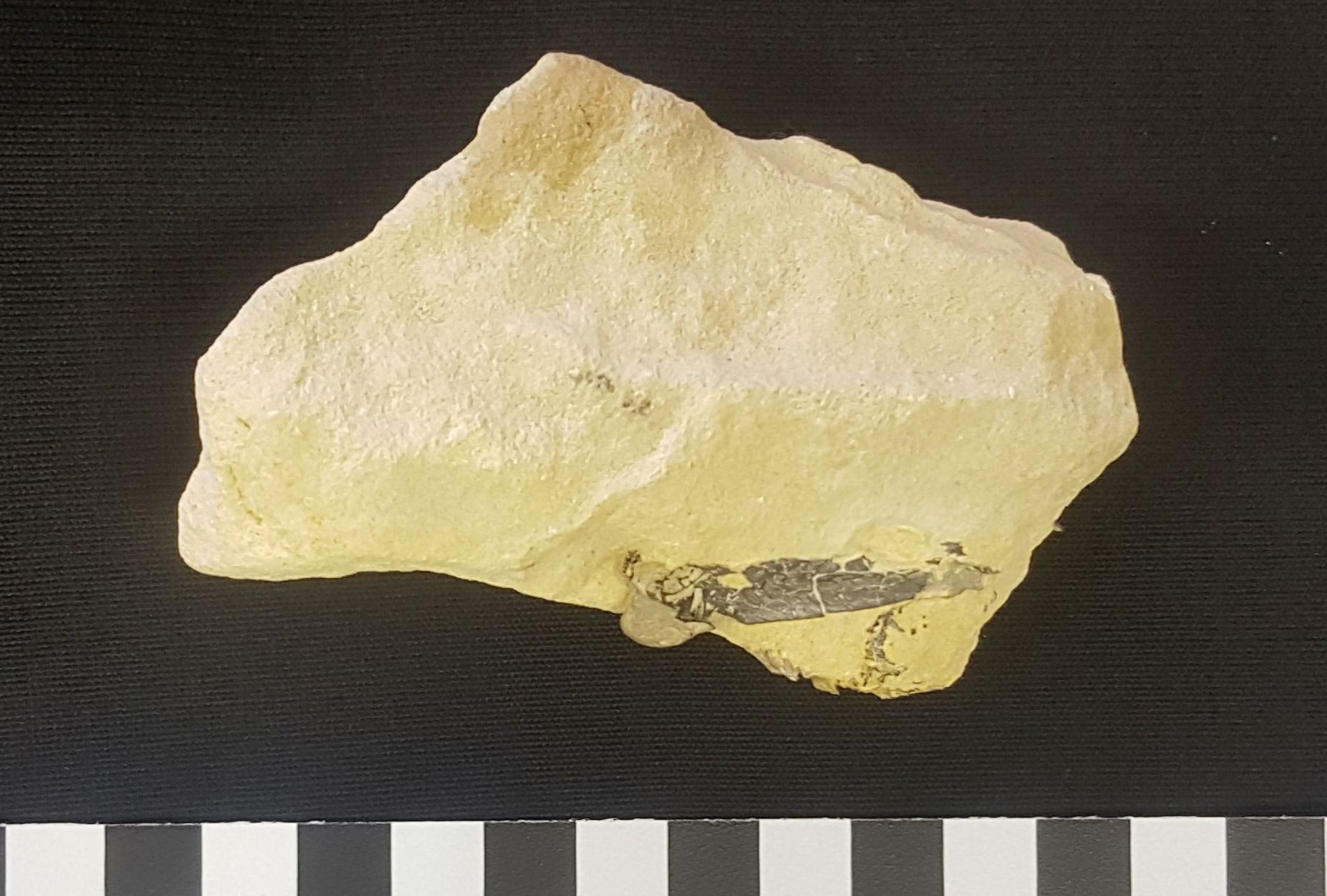
Unpainted and painted casts of 'Stan's' premaxillary bone
Unpainted and painted casts of 'Stan's' premaxillary bone
The bones are molded using silicone rubber. This
captures all the fine details. A resin polymer is poured into the molds to make the cast. The casts are then painted to make them look as close to the real bones as possible.
Scaling down
The Black Hills Institute CT-scanned 39 of Stan’s skull bones, including the braincase. Using this data they are able to produce a 3D-printed replica at 1:6 scale. The museum acquired a full set as part of Dr Hempel’s donation.

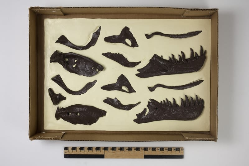
One of three boxes containing 1:6 scale casts of Stan's skull bones.
One of three boxes containing 1:6 scale casts of Stan's skull bones.

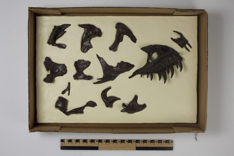
One of three boxes containing 1:6 scale casts of Stan's skull bones.
One of three boxes containing 1:6 scale casts of Stan's skull bones.

The 1:6 scale casts of Stan's skull bones
The 1:6 scale casts of Stan's skull bones
Over 30 complete replicas of Stan, each costing around $100,000, have been sold across the world. A bronze replica is on display outside Google’s headquarters in California. In the UK, Stan’s skeleton can be seen at The Manchester Museum and the Oxford University Museum of Natural History.
DR HEMPEL’S BEQUEST
Our collections team look after the two million objects in the museum. They also curate new collections, like Dr Hempel’s casts, before they can go on display.
Dr Andrew Hempel (1931-2017) was an eye surgeon
at Moorefields Hospital, London. His bequest of over 100 objects was photographed, conserved and catalogued by the museum’s collections team.

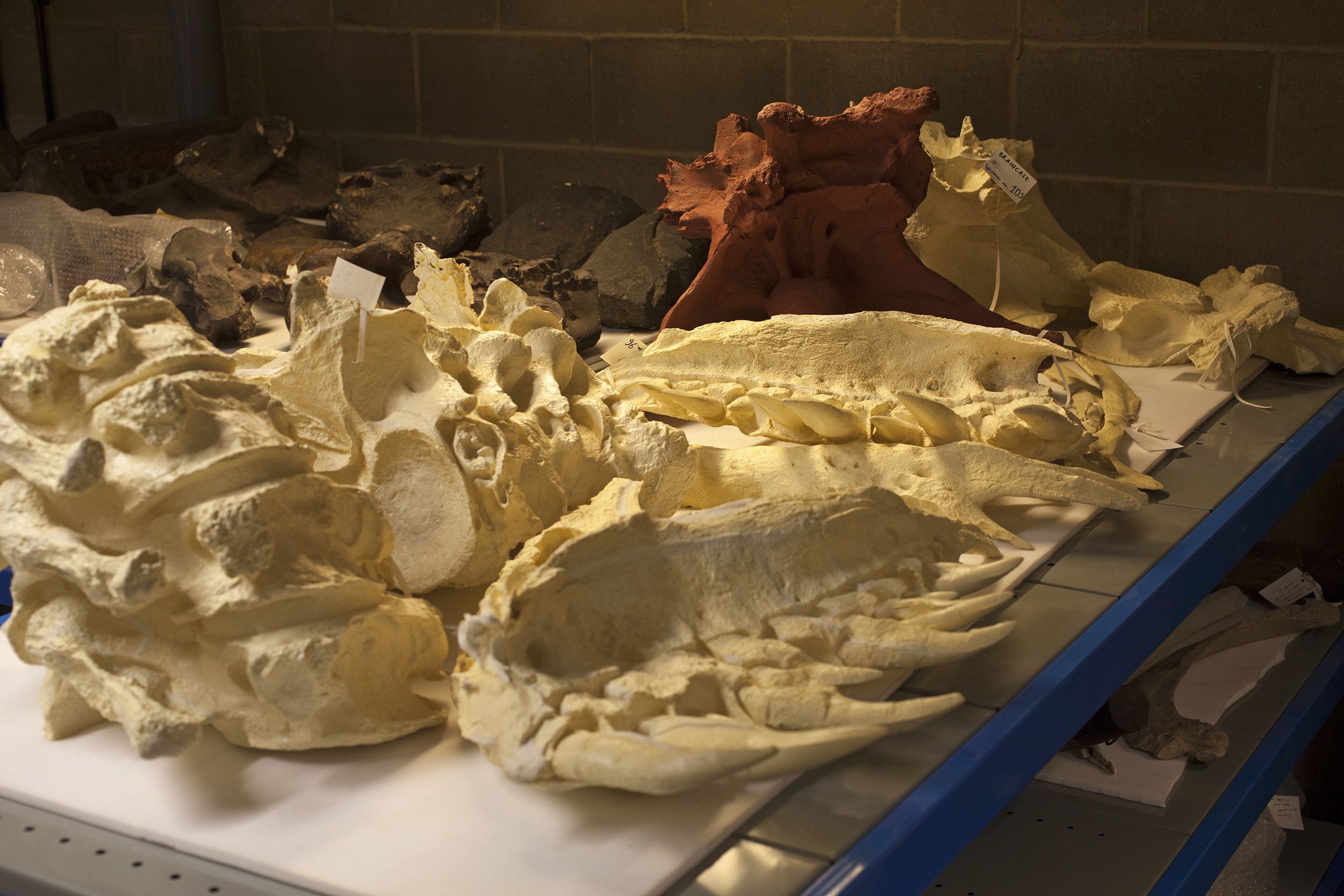
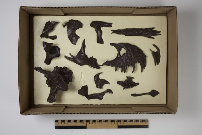
Unpainted casts of some of 'Stan's' skull bones in the museum store
Unpainted casts of some of 'Stan's' skull bones in the museum store
Dr Hempel’s collection is of great value to the museum and the Department of Earth Sciences at Cambridge. It will be useful for in teaching, research, display and outreach.

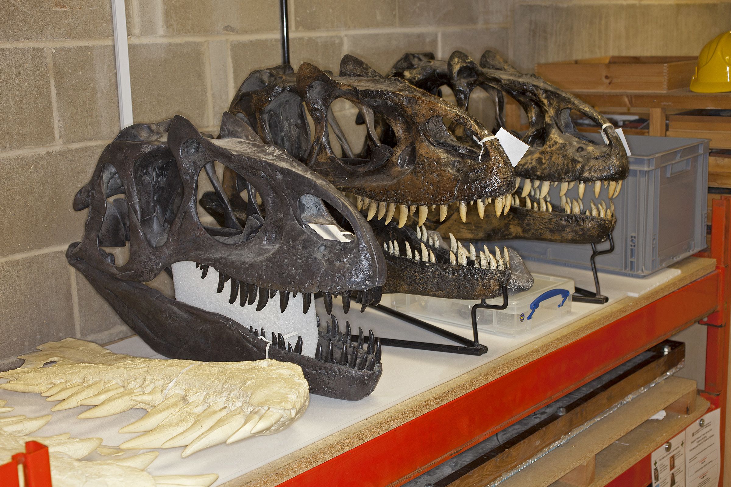
Three casts of an Allosaurus skull in the museum store
Three casts of an Allosaurus skull in the museum store
Object entry

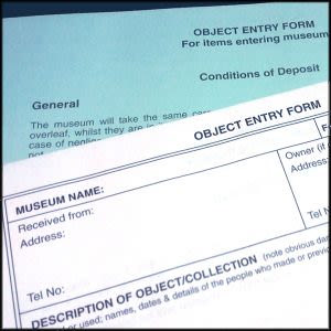
Museums must record the details of every object that comes into their care. Object entry forms are triplicate forms that many museums use. The white form is kept by the museum, the pink with the donor, and the blue is kept with the object.
Conservation
All the casts needed to be cleaned. Some also needed
repairing. The original skull bones are quite fragile, and the
replicas are the same. Damaged casts were repaired using a conservation grade adhesive called Paraloid-B72. Scuffs and scratches were repainted with matching colours.
Collections management systems

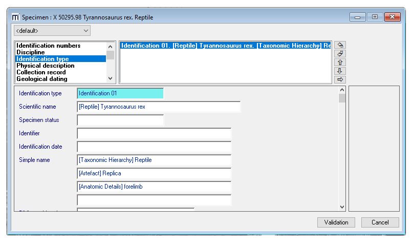
The details of each object are entered into the museum's electronic
database. Each has a unique number. The collections management system provides safe storage for this information and new details can be recorded. It also allows the collections team to track the location of objects if they are moved.
Storage

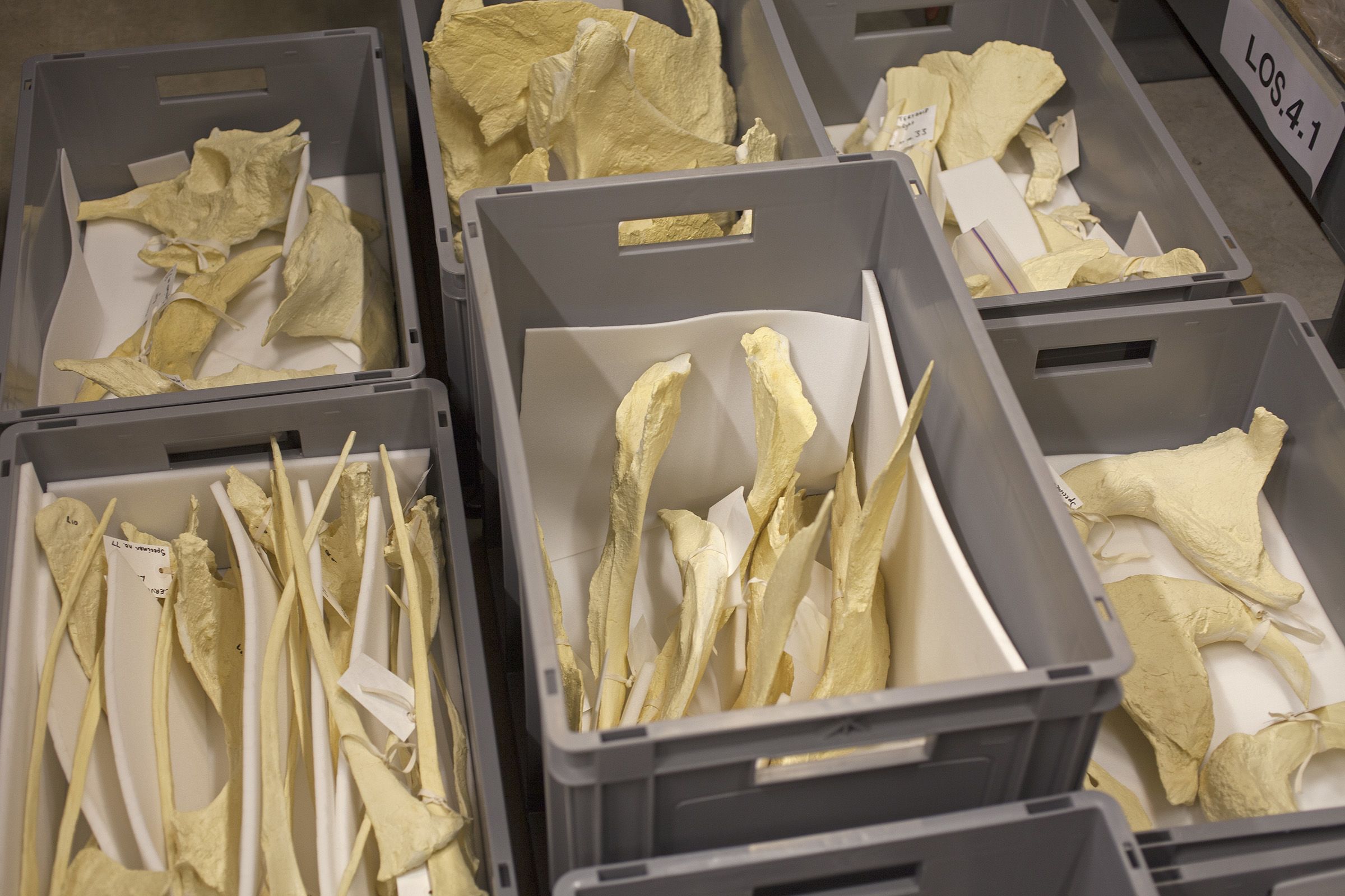
The collection is housed at the museum’s off-site store and laboratory. Most of the casts are large and have unusual shapes, which makes them a challenge to store. They are stored in stackable plastic crates. Each is lined with a polyethylene foam which is inert and acid-free to protect the specimens.
Labelling
Each object needs to be labelled with their own number.
They are attached with Paraloid-B72. It does not damage the objects and is also reversible. A fine-tipped permanent marker is used to write the numbers on each label. The labels are made from acid-free paper.
This online exhibition is an adaptation of a temporary display that we held in the museum between February 2018 and March 2019.


